Anatomy of a Hand – Hand Muscles
In this video lesson, you will discover the anatomy of a hand and including muscles of the human hand.
Enroll in the Drawing Academy Course
Pay once - Enjoy forever!
Only $297
Anatomy of a Hand
In this video part of the “Anatomy of a Hand” lesson, the same hand position is outlined in pencil next to the drawing of the hand bones, so you can cross-reference the relationship between hand’s bones and muscles.
The anatomy of a hand has two types of muscles:
– intrinsic
– and extrinsic
Extrinsic muscles of the hand take their origin from the lower part of the humerus, which is the long bone in the arm that runs from the shoulder to the elbow; as well as from the ulna and radius bones of the forearm.
Extrinsic muscles have long tendons, which insert into various carpal bones, metacarpal bones, and the phalanges of the hand.
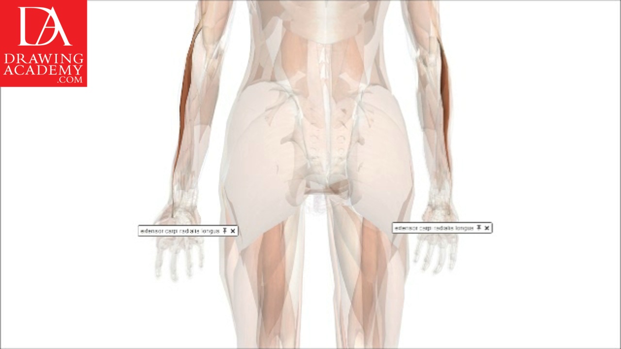
The human arm has more than 30 muscles from the elbow to the palm. These muscles and their tendons can be grouped into extensors and flexors:
1. Extensors are muscles located on the posterior side of the forearm and hand. Long extensors shape the back of the arm. These muscles supinate the palm or turn the palm upward and stretch back the wrist, fingers and thumb.
2. The second group is called the flexor muscles. They are located on the front of the forearm and form its shape. Flexor muscles pronate the palm or turn it down and bend the wrist, fingers and thumb. These muscles are used for gripping.
When it comes to the anatomy of a hand, unlike extrinsic muscles, which start from the arm, intrinsic muscles have origins and insertions directly on the hand. These muscles give mass to the hand, flexing, extending, adducting, and abducting the fingers and thumb.
Intrinsic muscles are divided into three groups:
1. The thenar group, which forms the thumb eminence, the thick oval-shaped mound at the thumb base. This group combines four muscles:
– adductor pollicis
– flexor pollicis brevis
– abductor policis brevis
– and opponens pollicis
2. The hypothenar group, which moves the little finger and forms the elongated mound of the palm between the wrist and the little finger. This group has three muscles:
– flexor digiti minimi brevis
– abductor digiti minimi
– and opponens digiti minimi
3. The interosseous group has eight muscles responsible for the adduction and abduction of the fingers. This group includes four palmar interosseous muscles and four dorsal interosseous muscles.
The extensor retinaculum holds the tendons of the extensor muscles in place. It is located on the back of the forearm, just next to the hand.
It continues with the palmar carpal ligament on the anterior (or internal) side of the forearm. It is a strong, fibrous band, which is thickened by the addition of many transverse fibres, and forms the dorsal carpal ligament.
The extensor digitorum is a muscle of the posterior forearm that extends the medial four digits of the hand. This is the dorsal transverse ligament which keeps the tendons together.
Then there is the two joint muscle called, extensor digiti minimi, which extends the wrist. This muscle moves the back of the hand toward the back of the forearm, and also extends the little finger, straightening it up from a fist position.
The extensor carpi ulnaris is a muscle located on the ulnar posterior side of the forearm. This muscle extends and adducts the wrist. When this muscle acts alone, it inclines the hand toward the ulnar side. Also it extends the elbow-joint.
The abductor digiti minimi muscles pulls the little finger away from the other fingers or abducts it. It plays an important role when we grasp large objects with outspread fingers.
Tendons of the hand are tied or bound against the bones by retinacula. Every retinaculum is not a part of any muscle but instead forms a band around tendons which holds it in place.
Tendons inserted within the fingers are bound to the knuckles.
Our fingers do not have any muscles. Apart from bones and tendons, the finger’s mass consists of fatty tissue, blood vessels and nerves. When it comes to the anatomy of a hand and palm anatomy in particular, the palm however has muscles.
The palm’s muscles are:
The interosseous muscles help to control the fingers. They are divided into two sets:
– (4) Dorsal interossei, which abduct the muscles away from the 3rd digit.
– (3) Palmar interossei, which adduct the muscles towards the 3rd digit.
The extensor carpi radialis brevis is a muscle in the forearm that extends and abducts the wrist. It is both an extensor and an abductor of the hand at the wrist joint. This muscle manipulates the wrist so that the hand moves away from the palm and toward the thumb.
The extensor carpi radialis longus is a muscle at the wrist joint that goes along the radial side of the arm. This muscle abducts the hand at the wrist, so it moves the hand toward the thumb.
Last are the extensor pollicis longus and extensor pollicis brevis. These muscles stretch the thumb. The extensor pollicis brevis both extends and abducts the thumb.
When drawing human hands and thinking about the anatomy of a hand, the fine artist must understand how a hand is attached to the forearm. The wrist bones are connected with the bones of the hand and they make one mass and as such, move together.
When it comes to the anatomy of a hand and its proportions, keep in mind that the wrist’s width is twice wider than its thickness. The wrist is thinner than the forearm bones at the place of connection, so there is a step down from the forearm bones to the wrist.
The wrist moves together with the hand on the forearm and also has some rotary movement. The twisting movement is provided by the forearm.
When it comes to the shape of the palm, the fine artist needs to keep in mind that it is slightly bowed from the knuckles to the wrist and from the first finger to the little finger and from side to side. The hand is also slightly arched on the back side from the first finger to the little finger. This arch is more prominent at the knuckles.
Also, when drawing hands applying a knowledge of the anatomy of a hand, remember, that the knuckle of the second finger is bigger and higher than the rest. The first and the little fingers have lower knuckles on the external sides.
The form of the hand at the little finger’s side is shaped by the abductor muscle. This bowed shape goes from the wrist to the middle of the first segment of the little finger.
When you draw a hand with an open palm and its fingers raised to the back side, make sure to depict the sharply raised tendons of the long extensors that are well prominent under the skin in such a position, this can be depicted from a memory when you know the anatomy of a hand.
When drawing the hand’s fingers, you need to remember both fingers’ proportions and the anatomy of a hand. As we already established, there are no muscles around the fingers’ bones. Finger bones have narrower shafts and wider ends. The finger joints are square; with the middle joint of each finger being the largest. The finger bones’ shafts also have square cross sections with rounded corners. The fingers have triangular tips.
When drawing hands by applying the anatomy of a hand, remember that from its back view, the fingers are slightly arched toward the middle finger.
When drawing a clenched fist, the thumb will overlap the middle finger.
The middle finger is the largest and longest, while the little finger is the smallest.
The little finger has the most freedom of movement compared to the other fingers.
Also, it is very important to remember the anatomy of a hand and proportional relationship between the hand and other parts of the body when drawing a human figure. The length from the wrist to the tip of the middle finger is the same as the length from the base of the base of the chin to the hairline above the forehead.
When the hand’s fingers are spread out so that the thumb in its furthest position from the tip of the middle finger, the distance between the tips of the thumb and the middle finger will be the same as the distance between the inner elbow depression and the wrist.
The width of four fingers is the same as the distance between the base of the chin and the root of the nose. It is also the same as the distance between the root of the nose and the eyebrow-line. And finally, the four fingers’ width measures the same as the distance between the eyebrows and the hairline.
Making life sketches is the best way to practice hand drawing and improving your knowledge of the anatomy of a hand. Drawing with the knowledge of a human’s hand anatomy will result in more realistic pictures.
