Neck Muscles – Anatomy for Fine Artists
In this video lesson, you will discover the muscle anatomy of the human body including the following parts:
– Neck Muscles
– Muscles of the Torso
– The Upper Limb Muscles
– The Lower Limbs Muscles
– Foot Muscles
Enroll in the Drawing Academy Course
Pay once - Enjoy forever!
Only $297
Neck Muscles – Anatomy of a Human Neck
In a separate video of the Drawing Academy Course I have presented the bones and muscles of the head and now we will examine the neck muscles.
The neck has a cylindrical shape, which follows the curve of the vertebral column.
It is always leaning slightly forward, even when the head is tilted backward.
The anterior (or front) part of the neck is rooted at the chest, while the posterior (or back side) of the neck is linked to the back.
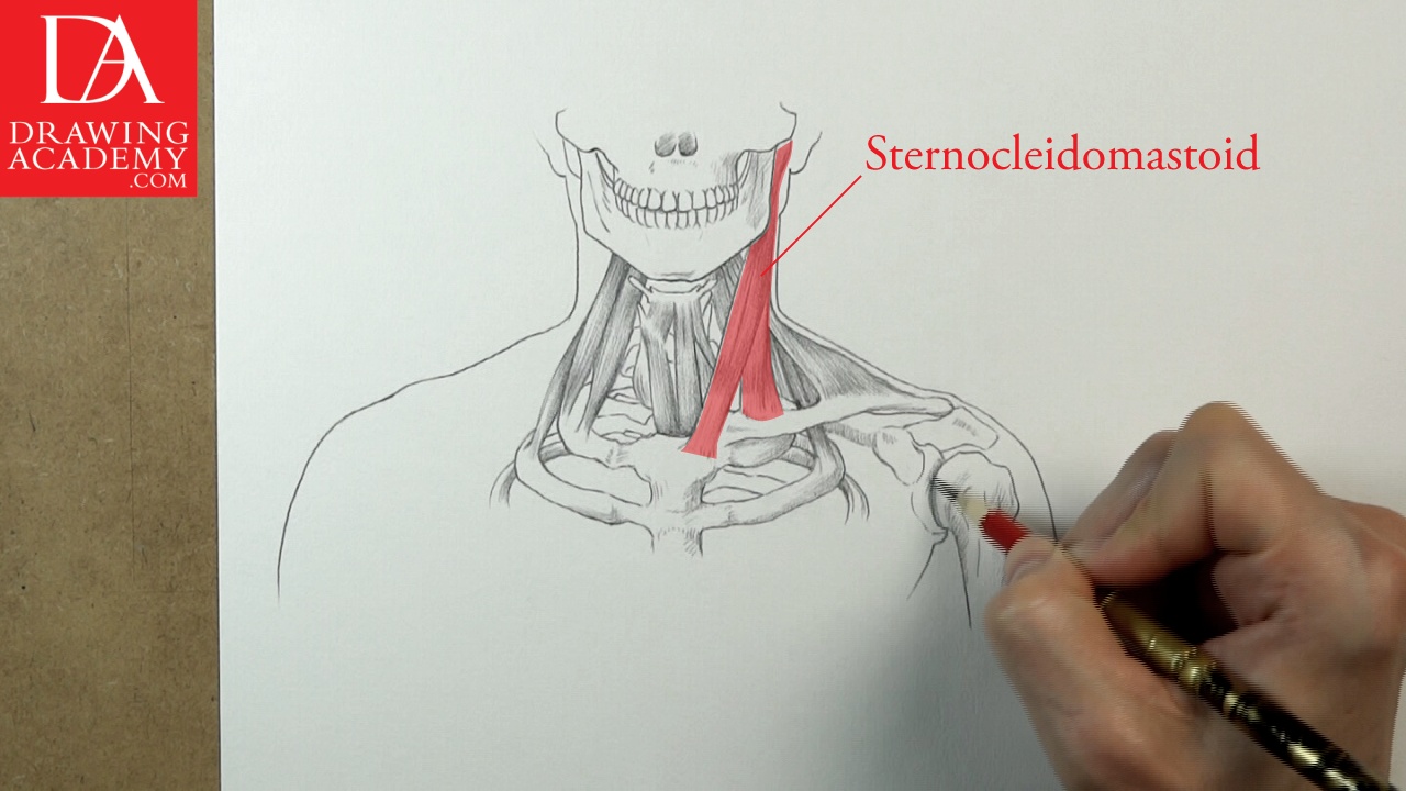
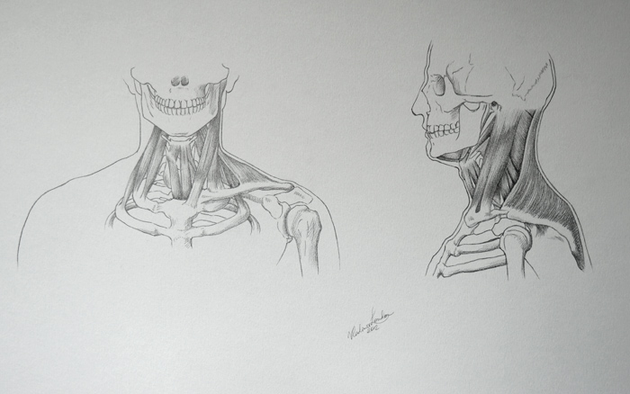
The sternocleidomastoid muscles are located on either side of the neck; however, for illustration purposes, I only presented one muscle on one side, so you can see what muscles are underneath on another side.
The Neck Muscles lying under the sternocleidomastoid muscle are as follows:
– Scalenus posterior
– Scalenus anterior
– Thyrohyoideus
– Sternothyroideu
– Omohyoideus
Let’s examine the sternocleidomastoid muscle because it is the closest to the surface of the neck, shapes the front of the neck and influences its appearance.
This muscle consists of two separate straps, which are attached to different bones – sternum and clavicle. That is why the sternocleidomastoid has two heads, which are called the sternal head and the clavicular head. The sternal head is also known as the sternal portion or medial head. The clavicular head also has two alternative names – the clavicular portion or lateral head.
The sternal head has a round cross-section, while the clavicular head is flat and broad. The small triangular space between the tendons of these two heads is called, the lesser supra-clavicular fossa. This space sometimes is noticeable on the neck surface and fine artists must be aware of this spot when making a model drawing with an exposed neck.
The space between the two tendons of the sternal heads on both sides of the neck is called the suprasternal notch, or the pit of the neck.
This spot is the important landmark for figurative fine artists, which helps to locate the beginning of the breastbone and its intersection with the collar bones, as well as serves as the pivot point of certain positions of the neck.
The sternal head tendons become more noticeable when the head is turned or tilted. The clavicular heads are less pronounced; they form the cylindrical surface of the neck.
The sternocleidomastoid Neck Muscles insert into the mastoid process of the cranium and base of the cranium behind the ears. Various actions of these Neck Muscles result in different movements of the head:
– The head flexion (or bending forward) happens when the Neck Muscles on both sides contract.
– Contraction of the posterior fibres on both sides causes the head to move up and back.
– The contraction of one side results in lateral flexion, making the head tilt to the shoulder.
– With the help of other muscles, the sternocleidomastoid muscles turn the head from side to side.
Other Neck Muscles include:
– Semispinalis capitis
– Splenius capitis
– Levator scapulae
– Scalenus posterior
– Scalenus anterior
– Trapezius
As you can see, the neck muscles are arranged in layers. They are strong and cover the back and sides of the neck. Neck muscles flex, extend, and rotate the head and also raise shoulders up.
On the front of the neck there are three prominent forms:
– Larynx (or voice-box)
– Cricoid cartilage
– Thyroid gland
The male larynx is bigger than the female’s, while women have a more prominent thyroid gland.
The structure of this part of the neck muscles as follows:
– Hyoid bone
– Thyrohyoid membrane
– Thyroid cartilage
– Laryngeal prominence
– Cricoid cartilage
– Thyroid gland
– Tracheal rings of trachea