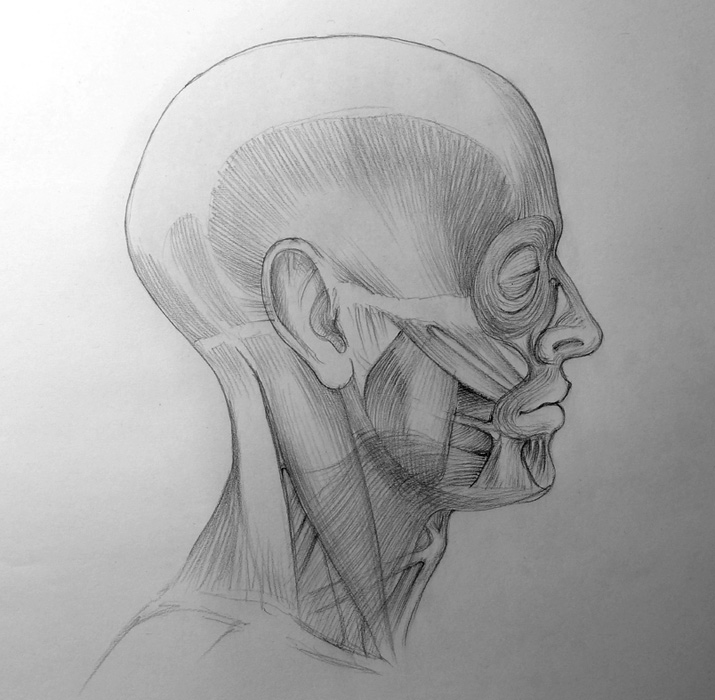
How to draw a head – Muscles Anatomy of the Skull
In this video lesson you will discover Anatomy of the Skull including muscles of the head, face and neck.
Enroll in the Drawing Academy Course
Pay once - Enjoy forever!
Only $297
Anatomy of the Skull
If your ambition is to become a proficient fine artist who can draw a portrait with confidence, then learning the Anatomy of the Skull is a must know topic.
Understanding the muscles of the face is very important when it comes to depicting facial expressions.
In Anatomy of the Skull, every muscle or group of muscles is responsible for a specific facial expression. Some expressions are done by one or two muscles, while others take several muscles at once. The facial muscles are not easy to see as they are covered by soft tissues, superficial fascia and fatty tissues. Nevertheless, the knowledge of human face anatomy becomes very handy when fine artists draw what they know about the Anatomy of the Skull rather than what they see.
Facial muscles are very special, as opposed to the skeletal muscles, which are attached from bone to bone; most of the facial muscles begin on a bone and insert into the skin, muscles, connective tissues or a combination of those. That is why when they contract, the skin and tissues are pulled, which changes the expression on a face and makes folds and wrinkles on the skin.
In the Anatomy of the Skull, the muscles most responsible for facial expressions are those in the eye and eyebrow area, as well as, the mouth.
The orbicularis oculi is a disk-shaped muscle. These muscles go around each eye socket or orbit.
The orbicularis oculi muscle consists of three parts:
• Orbital portion (which looks like a thick disk)
• Palpebral portion (which forms the eyelids)
• Lacrimal portion (which is not visible as it lies behind the eyes)
The orbital portion of the orbicularis oculi muscle is responsible for squinting, winking, and closing the eyes.
The orbicularis oris is the muscle, which goes around the mouth in Anatomy of the Skull. It also has a round shape, as reflected in its Latin name. There are a number of other muscles interwoven into the orbicularis oris and together are able to produce multiple expressions of the mouth. Contraction of the orbicularis oris causes shutting of the mouth or puckering of the lips. Multiple muscles around the mouth also performs other actions, which influence, mimic and contribute to the ability to speak.
The depressor anguli oris presses down the angle of the mouth. It is triangular in shape and has its origin on the lower border of the jawbone (or mandible) and inserts into the angle of the mouth. This is the muscle which depicts sadness or disapproval in the face. When the depressor anguli oris contract, they pull the corners of the mouth down.
Nearby is the depressor labii inferioris muscle. This muscle begins at the mandible, just under the depressor anguli oris and inserts into the lower lip’s skin and the lower area of the orbicularis oris muscle. When the depressor labii inferioris contracts, it pulls the lower lip downward, so usually the lower teeth become visible. It serves the purpose of speech and facial expressions.
Another muscles below the mouth is the mentalis, it is V-shaped and located on the chin. When this muscle contracts, it pull the skin on the chin upward, taking part in various expressions such as determination, anger, or disagreement.
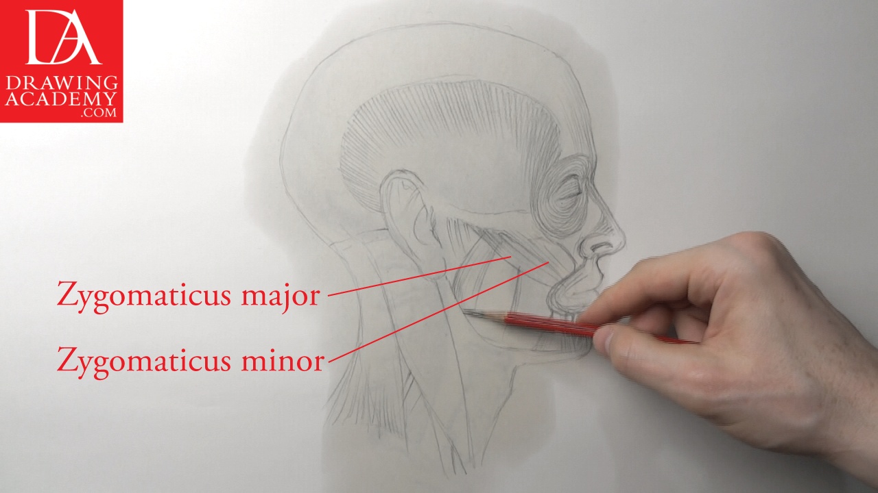
Another two muscles paired on both sides of the head are the zygomaticus major and zygomaticus minor. Both of these muscles start on the cheekbone. The zygomaticus major inserts into the corners of the mouth, while the zygomaticus minor goes into the upper lip’s skin. When these muscles contract, they pull the corners of the mouth and upper lip upward and outward; making a smiling expression on the face.
Another muscle that lifts up the upper lip is the levator labii superioris. It takes its origin on the lower border of orbit on the zygomatic and maxilla bones and inserts into the upper lip skin and orbital muscle of the mouth. When these muscles contract on both sides of the head, they pull the upper lip upward, usually exposing the upper teeth. The action of these muscles contributes to the facial expression of disgust or contempt. When only one side’s levator labii superioris is contracted, pulling only one side of the upper lip upward, it gives the sneering or snarling expression.
In the Anatomy of the Skull, the masseter muscle is essential for chewing. It is rectangular in shape; takes its origin on the zygomatic arch and inserts into the outside surface of the mandible and angle of mandible.
The primary function of the muscle is elevating the lower jawbone (or mandible), closing the mouth while chewing and clenching teeth. This muscle is also enables the lower jaw to move slightly forward. When the masseter is contracted, the superficial portion of this muscle can be seen as bulges on the cheeks, which could be also a sign of an emotionally tense expression.
Another muscle that assists the masseter in closing the lower jaw is called, temporalis. This is the widely spread flat muscle on the side of the temporal bone where it originates. Lower down, the temporalis converges into a tendon, which goes behind the zygomatic arch and has its insertion on the coronoid process and anterior ramus border of mandible.
When the front portion of the temporalis muscle, where fibres are located more vertically, contracts, it helps to move the lower jaw upward, closing the mouth. However, during contraction of the more horizontal fibres of the temporalis, the lower jaw moves backward from its neutral position.
Now let’s explore Anatomy of the Skull including muscles that depress the mandible or open the mouth. There are two separate muscles, which are called, mylohyoid and digastric. These muscles are located close and overlap each other in the under-plane of the jaw. The mylohyoid muscle starts from the inner surface of the body of mandible and inserts into a fibrous line that attaches from inside of the chin region of the mandible to the hyoid bone. The digastric muscle has two origins on both ends – first on the digastrics fossa on the lower inside of the mandible, and second on the mastoid notch of the cranium. It inserts on the hyoid bone by means of a tendon. These muscles are essential when it come to opening the mouth wide. They also elevate the hyoid bone when swallowing or speaking.
The buccinator is a horizontal muscle, located on both cheeks, forming their muscular walls. This muscle, when contracted, pulls lips backward horizontally, it also compresses the cheeks against the teeth. The buccinator controls the tension of the lips and therefore, often is called a “trumpeter’s” muscle. In facial expressions, it plays a role in making signals of annoyance, smirking and contempt.
The procerus muscle is located between the eyebrows on the lower central region of the forehead. It starts from the fascia of the nasal bone and the cartilage of the nose and inserts into the skin in the central region of the forehead. On either side of the procerus, there are two corrugators supercilii and depressor supercilii muscles. Together with the procerus, these muscles, when contracted, produce wrinkles near the root of the nose and depress inner ends of the eyelids. Facial expressions associated with the action of these muscles are anger, depression and intense concentration.
The frontalis muscle is located on the forehead (or frontal bone). It starts near the hairline and inserts into the skin and subcutaneous tissue of the eyebrow. The fibres this muscle interweaves with are the orbicularis oculi and the procerus muscles. The frontalis can produce three facial expressions with different meanings. When both sides of this muscle are contracted, the eyebrows are lifted up, producing horizontal wrinkles on the forehead, which can be reinterpreted as a sign of surprise disbelief. When only one side of the frontalis lifts, one eyebrow is raised, while the other eyebrow remains in its neutral position, such a sign can indicate an expression of bemusement. Not everyone can arch their eyebrow in this way. The third way the frontalis can contract is so the inner ends of the eyebrows are elevated, producing a sign of grief or sadness.
In the Anatomy of the Skull, the nose muscle is called, nasalis. It has two portions:
• The transverse portion
• The alar portio
The transverse portion has a fan shape and takes its origin on the maxilla near the lower outside of the nasal cavity. It goes upward, covering the side of the nose, and inserts on the fascia of the top plane of the nose. The alar portion takes its origin on the maxilla below the nasal cavity, and inserts into the skin of the nose and lower margin of the nostril. The visible action of the nasalis is very subtle and doesn’t contribute to any facial expressions.
Another muscle in the nose region is the levator labii superioris alaeque nasi. It’s quite a long name for a relatively short muscle; its name can be abbreviated as LLSAN. This muscle starts from the maxilla near the root of the nose and inner corner of the eye and goes downward where it inserts into the wing of the nose, the skin of the upper lip and the skin of the nasolabial furrow. By contracting, this muscle lifts up the wing of the nose and the middle part of the upper lip together with the nasolabial furrow, or the crease that goes from the wing of the nose downward to the side of the mouth; it is also creates various wrinkles on the side of the nose. This action usually is associated with the sign of disgust.
According to the Anatomy of the Skull, the sternocleidomastoid muscle consists of two parts:
• The Sternal head (also known as the sternal portion or medial head)
• The Clavicular head (also called the clavicular portion or lateral head)
The sternal head has its origin on the sternum and the clavicular head starts on the clavicle. These muscles insert into the mastoid process of the cranium and superior nuchal line of the occipital bone (or base of the cranium).
The following muscles in Anatomy of the Skull are responsible for bending the head into different directions:
• The head moves forward when both sides of these muscles contract. This movement is called, flexion.
• Contraction of only posterior fibres of both sides moves the head up and backward.
• Contraction of one side tilts the head closer to the shoulder on the side of the contracting muscle. This movement is called, lateral flexion.
• With the help of other muscles, the sternocleidomastoid rotates the head from side to side.
When drawing the neck, these muscles are pronounced under the skin and need to be observed and depicted to make a realistic portrait.
In this video part of the Anatomy of the Skull, we explored the head, face and neck muscles and their actions, as well as, what facial expressions are caused by certain facial muscles. This is a very brief overview and I would suggest you read a book on this topic to deepen your human head anatomy knowledge.
Also, when it comes to reading and understanding signs of body-language, you may refer to my book, BODY LANGUAGE IN FINE ART: How to Read the Old Masters’ Paintings – Secrets of Body Language In Figurative Fine Art.
This book is a useful resource for fine artists and fine art lovers who want to discover hidden messages left by the Old Masters that can only be understood by reading a non-verbal language depicted in oil paintings, drawings and sculptures.