Human anatomy for fine artists – Leg Bones
In this video part, you will discover the Leg Bones anatomy including the pelvis – which is part of the torso – as well as the bones of the leg.
Enroll in the Drawing Academy Course
Pay once - Enjoy forever!
Only $297
Pelvis Anatomy
Let’s begin with the pelvis.
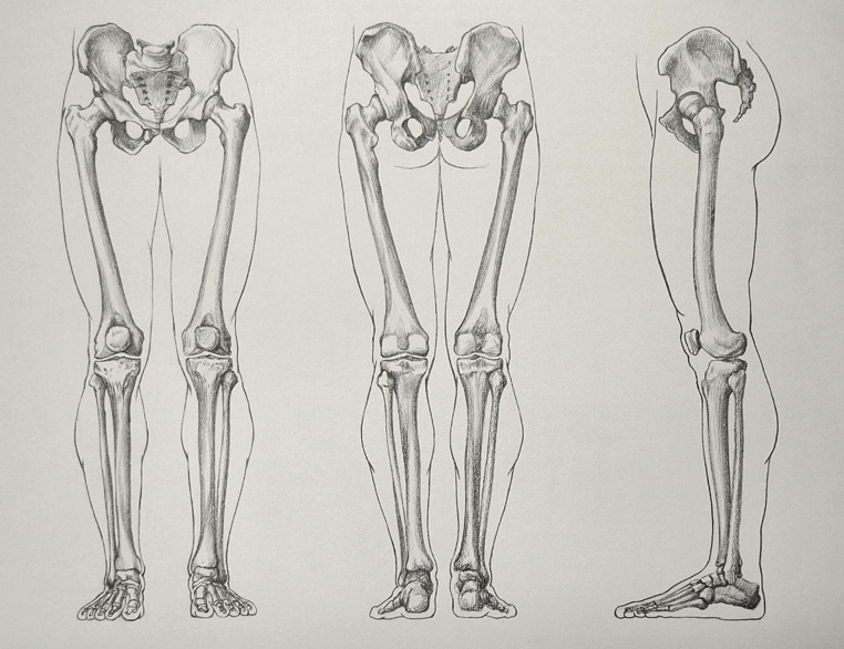
The pelvis is the bony structure between the legs and the torso. It connects the spine to the thigh bones.
An adult’s pelvis is constructed of two hip bones, on both left and right sides of the body. In childhood, each of two bones of the pelvis consists of three separate bones, which are fused together during puberty. These three sections are:
– The Ilium
– The Ischium
– The Pubis
Together with the Sacrum, these bones form the pelvis. These bones form a single unit and do not move independently of each other.
The area enclosed by the pelvic bones is the pelvic cavity. This section lies below the abdomen and mainly consists of the reproductive organs and the rectum.
Apart from connecting the upper part of the body with the leg bones, the pelvis also bears the weight of the upper body when sitting, standing or walking. It is also serves as the structure to which the muscles responsible for body movement and posture are attached. Therefore, the pelvis has strong and rigid bones.
The two ilium bones of leg bones resemble a bowl shape, or basin, where the pelvis name comes from. The pelvis is Latin for basin. In fact, one of the functions of the pelvis is to support the internal organs from underneath, as if they were placed into a basin.
This is very different from animals which walk on four limbs, whose pelvises do not support their internal organs as the human pelvis does.
The top edge of the ilium bone is called the iliac crest. The location of the iliac crest is noticeable on athletically shaped figures.
The front end of the iliac crest has a bump, which is called the anterior superior iliac spine (or ASIS). The two ASIS protrusions are important points of the pelvis, which helps fine artists to determine and depict the pelvis tilt. For example, this marble figure has a well-pronounced pelvis tilt because it rests most of its bodyweight on its right leg, while its left leg is bent at the knee.
Anatomically, the male pelvis and the female pelvis have a similar structure but differ from each other in proportions and shapes.
In general, the female pelvis has a relatively wider hip span while the male pelvis is narrower. The male pelvic brim has a heart shape, while the female pelvic’s brim has a wider and more rounded shape – like an oval.
The pubic angle of the male pelvis has a narrow triangle shape, while the female’s pubic angle is wider.
The male pelvis is more upright, while the female’s pelvis is tilted forward. The female sacrum is shorter and wider and has more space between the sacrum and pelvis; while the male’s sacrum is longer and narrower and located closer to the pelvis.
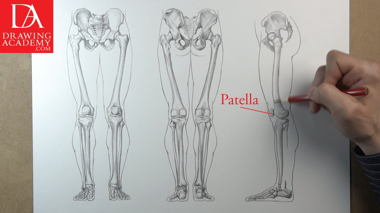
Now let’s examine the leg bones.
The leg bones are constructed similar to the upper and lower arm. Like the single bone in the upper arm, there is only one bone in the upper leg – a single bone called the femur.
The lower leg is in fact like the lower arm, which has two bones; these bones are called the tibia and the fibula.
The upper part of the femur has two bony projections:
– The greater trochanter
– The lesser trochanter
The greater trochanter comes quite close to the body surface and plays an important role in model drawing. Fine artists must be able to locate this bony part to depict realistic images of models.
The upper leg bone ends with the cylindrical neck of the femur and the spherical head of the femur, which fits into the hip socket of the pelvis.
At the lower end of the femur, there are two bony projections, called the lateral condyle of femur and the medial condyle of femur.
The leg bones are divided into three parts:
– The thigh – which extends from the pelvis to the knee
– The leg – which goes from the knee to the foot
– The foot
The femur, or the thigh bone is the biggest, strongest, and heaviest bone in the human body. At the knee, the femur bone rests on the tibia, or shin bone, forming the hinge joints between these bones.
The upper part of the tibia has two protrusions:
– The lateral condyle of tibia
– The medial condyle of tibia
Together with the lateral and medial conyles of femur, these bony parts are very close to the body surface and form the shape of the knee area.
The fibula is more slender than the tibia. This bone has the following parts:
– The head of fibula
– The shaft of fibula
– The lateral malleolus of fibula – found at the lower end
The head of fibula can be detected on the side of the leg at the knee area. Fine artists must recognize and depict this anatomical feature of the leg.
The knowledge of the knee’s anatomy is very important for a figurative fine artist. Many artists oversimplify the knee region when making drawings of the leg. To avoid this amateurish mistake, let’s examine the knee’s construction.
The knee joint is the largest synovial joint in the human body. A synovial joint is one in which the ends of the bones are enclosed in a capsule containing a thick, slippery liquid called synovial fluid. The capsule is made of strong, fibrous tissue and is lined with a membrane called the synovial membrane. The bone ends are covered in a smooth layer of a tough, rubbery substance known as cartilage.
Let’s take a closer look at the knee joint. It includes the condyles of the femur and condyles of the tibia. In the extended leg, the triangular bone, called the patella (or kneecap) is positioned just in front of the condyles of the femur.
The patella is quite small but plays a significant role in the shaping of the knee’s form.