Muscles of the Leg – Anatomy for Fine Artists
Video Lesson Description
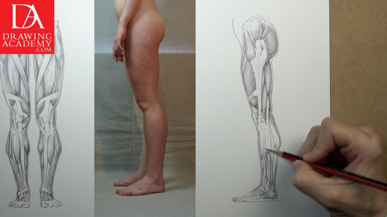
In this video part, you will discover the main Muscles of the Leg including the upper and lower leg muscles and tendons.

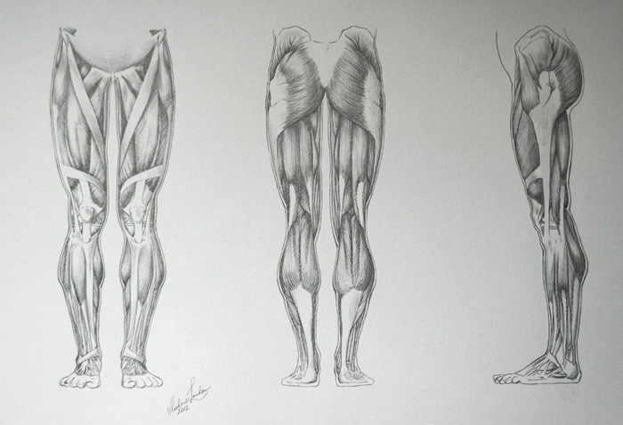
Muscles of the Leg
Let’s start with the muscles of the upper leg. The thigh can be divided into three compartments or groups:
– The anterior compartment
– The posterior compartment
– The medial compartment
The anterior compartment of the thigh contains the quadriceps or extensor group, which includes the following muscles:
– Vastus lateralis
– Vastus medialis
– Rectus femoris
– Vastus intermedius – which is covered by the rectus femoris (not visible on this drawing)
Vastus lateralis, vastus medialis and vastus intermedius all have their origin on the femur, or thigh bone. The fourth muscle of this group, rectus femoris, originates on the pelvis. All four muscles have a common tendon that inserts into the patella or kneecap and patellar ligament.
The powerful quadriceps group has the main action of extending the lower leg, straightening it up from being bent in the knee position.
Another muscle of the upper leg that doesn’t belong to any group is called, sartorius. This muscle is the longest in the human body and shaped like a strap. It originates near the bony protrusion of the pelvis, called the anterior superior iliac spine, and inserts into the medial surface below the medial condyle of tibia.
The sartorius helps the other muscles of the leg to bring the lower leg into a cross-legged position.
The medial compartment of the thigh contains the adductor group, which consists of the following muscles:
– Adductor mangus (not visible on this drawing)
– Adductor longus
– Adductor brevis
– Pectineus
– Gracilis
The adductor mangus is the largest muscle of the group. Despite that it is located deep inside the thigh and completely hidden by other muscles, the adductor mangus contributes to the shape of the inner thigh.
The muscles of the adductor group are mainly responsible for the adduction of the upper leg at the hip joint and in such movements as, for example, when holding the knees tightly together. In addition, the adductor longus, the pectineus, and the adductor mangus also help to flex the upper leg at the hip joint, bringing the knee upward.
Now, let’s have a look on the muscles of the leg of the posterior side of the leg.
The posterior compartment of the thigh containing the hamstring or flexor group, includes the following three muscles:
– Biceps femoris
– Semitendinosus
– Semimembranosus
All three muscles begin on the pelvis and their origin is concealed by the gluteus maximus. These flexor muscles insert on the upper portions of the fibula and tibia – the bones of the lower leg.
The hamstring group of muscles has the main action of extending the thigh, straightening it up, and flexing or bending the leg at the knee joint. In addition, the semitendinosus and the semimembranosus muscles also rotate the tibia on the femur when the knee is bent. The biceps femoris is responsible for the lateral rotation of the tibia when the knee is flexed.
The third group of the upper leg is called the gluteal group. It contains the following muscles:
– Gluteus maximus
– Gluteus medius
– Tensor fascia latae
– Gluteus minimus – which is completely covered by the gluteus medius muscle
These four muscles of the leg cover most of the pelvis from the lateral and posterior sides. The gluteus maximum and tensor fascia merge into the iliotibial tract, which goes down into the lateral side of the upper leg and inserts into tibia bone of the lower leg.
As evident from its name, the gluteus maximus is the largest and most powerful muscle of the group. It is responsible for extending (moving backward) the thigh and also helps in the lateral rotation of the thigh, which is the rotation of the upper leg outward. Together with the iliotibial tract, this muscle helps to maintain the upright posture of the human body.
The gluteus medius contributes to the abduction of the thigh, which is moving the upper leg sideways, away from the body. It also helps rotate, medially, the upper leg.
The tensor fascia latae helps abduct the upper leg by rotating it slightly inward.
Now, let’s examine the muscles of the lower leg.
There are three compartments of the lower leg:
– Anterior compartment
– Posterior compartment
– Lateral compartment
The anterior compartment contains the exterior group, which includes the following muscles of the leg:
– Tibialis anterior
– Extensor digitorium longus
– Extensor hallucis longus
The tibialis anterior muscle begins on the lateral surface of upper shaft and the lateral condyle of the tibia and inserts into the medial cuneiform and the base of first metatarsal of the foot. The tendon of this muscle is noticeable in the ankle region when the foot is raised up.
The main action of the tibialis anterior is to assist in lifting up the front part of the foot. This movement is called, dorsiflexion. This muscle also helps in the movement called, inversion, when the sole of the foot is being turned toward the medial line of the body.
The extensor digitorium longus begins from the lateral condyle of tibia, head and anterior surface of fibula, and the interosseous membrane. It inserts into the middle and distal phalanges of the 2nd to the 5th toes.
This muscle extends or lifts up the toes of the foot. Its four tendons become apparent on the foot surface when the toes are raised. The extensor digitorium longus also helps to lift up the front part of the foot contributing to the dorsiflexion of the foot.
The extensor hallucis longus takes its origin on the medial surface of fibula and the interosseous membrane, which is located between the bones of the lower leg. This muscle inserts into the distal phalanx of the great toe.
The main action of the extensor hallucis longus is to extend the great toe, lifting it upward. The muscle tendon becomes noticeable on the surface of the foot during this action. This muscle also contributes to the dorsiflexion movement.
The posterior compartment contains a flexor group, which includes:
– Gastrocnemius
– Soleus muscles
These two muscles of the leg are sometimes called the triceps surae, because together they have three heads, which start from the femur, the fibula and the tibia bones, and one common tendon, which inserts into the calcaneus or heel bone. Most of the mass of these muscles is located on the upper half of the lower leg.
This muscle group flexes the foot downward at the ankle joint and lifts the heel up from the ground; this movement is called the plantar flexion. The gastrocnemius muscle also helps to flex the lower leg at the knee joint.
The lateral compartment of the lower leg contains the peroneal group, which consists of:
– Peroneus longus
– Peroneus brevis muscles
Both muscles of the leg take their origin on the fibula bone, and inserts into different points of the foot.
The peroneal group of muscles helps to move the foot downward, contributing to the plantar flexion, as well as, assists in eversion action when moving the foot outward.
To find out more on the lower limb muscle’ anatomy you may refer to an anatomy book for fine artists.
- Receive 15 new videos monthly (45 in total)
- Incredible discount – $4,164
- Bonuses - Fine Art eBooks and Videos
- Drawing Academy Diploma of Excellence after course completion in 3 months
- Personal coaching by Drawing Academy Tutors
- Lifetime membership. Free after the 3rd month

- Immediate access to all 45 video lessons
- Incredible discount – $4,198
- Bonuses - Fine Art eBooks and Videos
- Drawing Academy Diploma of Excellence after course completion in 3 months
- Personal coaching by Drawing Academy Tutors
- Lifetime membership. No more payments