Foot Bones – Human anatomy for fine artists
Video Lesson Description
In this video part, you will discover the complex structure of the foot bones, which a figurative fine artist must know in order to depict a foot realistically.

Foot Bones
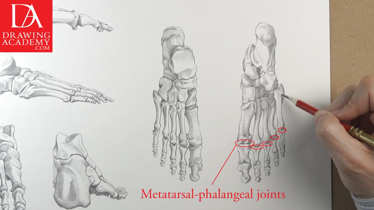
Every foot consists of 26 foot bones. The foot has a quite complex structure, which reminds one of the arrangement of the hand bones. Like the hand, it has 14 phalanges, located in the toes, and 5 metatarsal bones, which can be compared to the metacarpal bones of the hand.
The largest tarsal bone is the calcaneus, which is also known as the heel bone.
The heel bone has a chunky posterior projection, called the calcaneal tuberosity, which forms the prominence of the heel. The prominence of the heel is the place where the calcaneal tendon is attached, which is also known as the Achilles tendon. This tendon belongs to the gastrocnemius and soleus muscles.
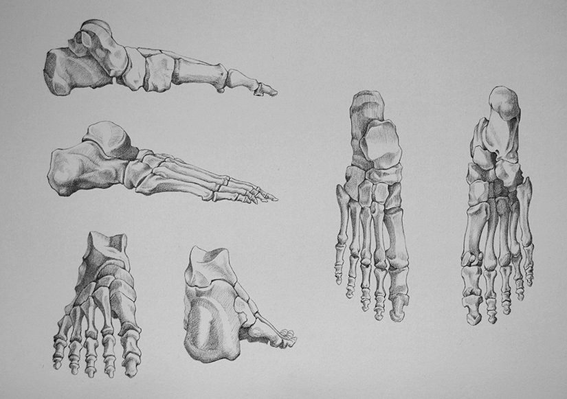
The talus is the only tarsal bone of the foot bones that has no muscular attachments.
Within the foot bones, both the calcaneus and the talus bones form the proximal row of the tarsals.
The remaining 5 tarsal foot bones are:
– The navicular
– First cuneiform (or medial cuneiform)
– Second cuneiform (or middle cuneiform)
– Third cuneiform (or lateral cuneiform)
– The cuboid
Together, these foot bones form the distal row of the tarsals. Because these five tarsal foot bones are positioned in front of the calcaneus and talus, they are also referred to as anterior tarsal bones.
Various bands and ligaments connect and keep the tarsal foot bones together. Tarsal foot bones have flat or plane joints, which allow a slight gliding movement against each other, which is very helpful when the foot is placed on rough terrain.
There are five metatarsal foot bones. They have elongated shapes and start from the anterior tarsal bones. These bones are not placed flat; instead they form the transverse arch, which helps to absorb shock during walking, running and jumping.
The transverse arch is one of the three main arches of the foot. The other two arches are:
– The medial longitudinal arch
– The lateral longitudinal arch
The first metatarsal bone is the shortest, thickest and plays an important role in mobility. It also provides attachment for several tendons. The second, third, and fourth metatarsal foot bones are the most stable of the metatarsals. They are well protected and have only minor tendon attachments and are not subjected to strong pulling forces.
Apart the five metatarsal foot bones, the forefoot includes the phalanges as well.
From the second to the fifth, lesser toes have three phalanges per toe:
– Proximal (first) phalanx
– Middle (second) phalanx
– Distal (third) phalanx
The great toe has only phalanges:
– Proximal phalanx
– Distal phalanx
The foot is designed to bear the heavy weight of the whole body. Its construction is arranged in a series of arches, which converge on the heel. The foot bones form a transverse arch. The inner arches of the foot are successively higher, forming half of the transverse arch, whose completion is in the opposite foot. Opening gradually toward the ankle, this arching movement finally culminates in the two columns of the leg and the arch between; wherefore the leg is placed somewhat to the inside of the central line of the foot.
In all positions, the foot tends to keep itself flat with the ground; the arches of the foot changing accordingly. In action, the foot comes almost into a straight line with the leg, but when settling upon the ground, the outer or heel side strikes first and the whole foot settles toward the inside.
The knowledge of the human skeleton bones is essential for realistic figurative drawing.
To find out more about human anatomy, you may refer to anatomy books for fine artists.
- Receive 15 new videos monthly (45 in total)
- Incredible discount – $4,164
- Bonuses - Fine Art eBooks and Videos
- Drawing Academy Diploma of Excellence after course completion in 3 months
- Personal coaching by Drawing Academy Tutors
- Lifetime membership. Free after the 3rd month

- Immediate access to all 45 video lessons
- Incredible discount – $4,198
- Bonuses - Fine Art eBooks and Videos
- Drawing Academy Diploma of Excellence after course completion in 3 months
- Personal coaching by Drawing Academy Tutors
- Lifetime membership. No more payments